Osteochondral lesions
Osteochondral lesions of the talus (OCLT) are a type of injury that affects the cartilage and underlying bone of the talus, a bone in the ankle joint. While this condition is relatively uncommon, it can lead to long-term pain and mobility issues if not treated properly. If you’re experiencing ankle pain or difficulty with movement, it might be related to an osteochondral lesion of the talus. Here’s what you need to know about this condition.
What is an Osteochondral Lesion of the Talus?
An osteochondral lesion of the talus refers to damage to both the cartilage and bone of the talus. The talus is a bone that, along with the tibia and fibula, forms the ankle joint.
The cartilage lining the talus is essential for smooth movement in the ankle joint. Any damage can result in pain, swelling, stiffness, and difficulty bearing weight on the affected ankle.
Causes of Osteochondral Lesions of the Talus

OCLTs are often caused by trauma to the ankle, but there are a variety of factors that can contribute to their development:
- Ankle Sprains or Fractures
- Repetitive Stress
- Poor Joint Alignment
- Degenerative Conditions
Symptoms of an Osteochondral Lesion of the Talus
The symptoms of an osteochondral lesion of the talus can vary depending on the severity of the injury. Common signs include:
- Pain in the ankle, especially when bearing weight or during movement.
- Swelling around the affected ankle joint.
- Stiffness or a feeling of “locking” in the ankle, making it difficult to fully move the joint.
- Tenderness when the affected area is pressed.
- Instability or a sensation that the ankle is weak, leading to a higher risk of reinjury.
- Grinding or popping sensations in the ankle during movement (in more severe cases).
How Are Osteochondral Lesions of the Talus Diagnosed?

Our doctors typically begin by performing a physical examination of the ankle to assess pain, swelling, and range of motion. Imaging tests are necessary to confirm the presence of an osteochondral lesion and determine its size and severity. These tests may include:
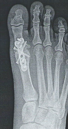
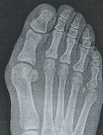
- X-rays to check for fractures or other bone-related issues.
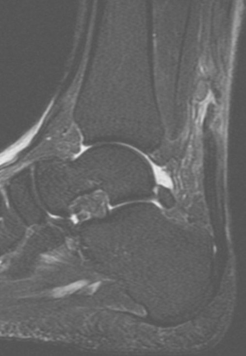
- MRI (Magnetic Resonance Imaging) to get a detailed image of the cartilage and soft tissues, helping to identify the exact location and extent of the lesion.
- CT (Computed Tomography) Scan can also be used in some cases to provide a more detailed view of the bone structure.
Treatment Options for Osteochondral Lesions of the Talus

Treatment for osteochondral lesions depends on the severity of the lesion, the symptoms, and the individual’s activity level. Treatment options can be divided into non-surgical and surgical approaches:
Non-Surgical Treatment
For mild to moderate lesions, non-surgical treatment may be effective in managing symptoms and promoting healing:
- Rest and Activity Modification
- Physical Therapy
- Nonsteroidal Anti-Inflammatory Drugs (NSAIDs)
- Bracing or Orthotics
- Cortisone Injection
- Regenerative Therapies
Surgical Treatment
In cases where non-surgical treatments do not provide relief or if the lesion is severe, surgery may be necessary. Surgical options include:

- Arthroscopy
This minimally invasive procedure involves inserting a small camera (arthroscope) into the ankle joint to visualize and remove damaged tissue or cartilage. In some cases, our doctors may also smooth out rough cartilage or repair damaged areas. - Microfracture Surgery
This procedure involves creating tiny holes in the bone beneath the damaged cartilage to stimulate the growth of new cartilage. While this can be effective in promoting cartilage repair, it may take time to see full results. - Cartilage Replacement
If the cartilage damage is extensive, our doctor may recommend using synthetic cartilage to replace the damaged area. - Osteochondral Autograft Transplantation (OATS)
In cases with severe damage, this technique involves transplanting healthy cartilage to replace the damaged area.
Recovery and Outlook
Recovery from an osteochondral lesion depends on the severity of the injury and the type of treatment used. Non-surgical treatments may involve weeks to months of rest and physical therapy. Recovery from surgery typically takes several months, with a gradual return to physical activity.
In general, people with mild to moderate lesions can expect a good outcome and a return to normal activity levels.
Conclusion
Osteochondral lesions of the talus can lead to significant pain and discomfort, but with appropriate treatment, you can return to your regular activities. Whether treated with non-surgical methods or through more advanced surgical options, there are effective ways to address the injury. Contact our office today for an accurate diagnosis and discovering the right treatment plan.
Frequently Asked Questions
X-rays are highly recommended in order for our doctors to rule out fractures and other injuries.
A MRI or CT is not required initially however, will likely be recommended if there is suspicion for an OCLT.
Yes, untreated or severe lesions can lead to the development of osteoarthritis in the ankle joint over time. Early intervention is key to minimizing this risk.
Returning to sports or high-impact activities will depend on the severity of the lesion and the type of treatment. After recovery, many people can return to normal activity, but the timeline and intensity of activity will vary.
Small lesions with minimal cartilage damage may heal on their own. However, larger lesions, often require surgical intervention to restore joint function and prevent long-term complications.
Ready to schedule an appointment or have questions for our staff?
Contact our office or click the link below to schedule an appointment.